What Is an OCT Eye Exam? Complete Guide for Patients and Clinics in Bosnia and Herzegovina (2025)
An OCT eye exam is one of the most important diagnostic imaging tests in modern ophthalmology. This quick, painless and non-invasive procedure provides a highly detailed view of the retina and optic nerve in microscopic resolution. In this guide, we explain everything patients and clinics need to know about the OCT eye exam — what it is, how it works, when it is performed, and what it reveals.
What Is an OCT Eye Exam and How Does It Work?
OCT (Optical Coherence Tomography) is a non-invasive imaging method that uses light waves to generate cross-sectional images of the retina. It can best be described as “an ultrasound that uses light instead of sound,” but with significantly higher resolution.
An OCT eye exam allows:
-
precise macular evaluation
-
glaucoma assessment through RNFL and GCC analysis
-
detection of edema, membranes and degenerative changes
-
follow-up of treatment effects (e.g., anti-VEGF therapy)
For a professional explanation of OCT technology, you can refer to the American Academy of Ophthalmology:
https://www.aao.org/eye-health/treatments/what-is-oct
Why Is an OCT Eye Exam Performed? (Most Common Indications)
An OCT eye exam is essential in diagnosing:
-
age-related macular degeneration (AMD)
-
diabetic macular edema (DME)
-
glaucoma
-
epiretinal membranes (ERM)
-
retinal tears and detachments
-
high myopia
-
post-operative monitoring
OCT provides information that no other imaging method can offer.
Does an OCT Eye Exam Hurt and Do Your Pupils Need to Be Dilated?
An OCT eye exam is:
-
completely painless
-
does not touch the eye
-
usually lasts 10–30 seconds
In most cases, pupil dilation is not required unless the patient has extremely narrow pupils or significant lens opacities. A regular OCT eye exam is especially important in glaucoma, diabetes and macular diseases.
Macular OCT – What Does It Show?
The macula is the central part of the retina responsible for sharp and detailed vision. An OCT eye exam of the macula shows:
-
macular thickness
-
presence of edema
-
subretinal fluid
-
epiretinal membranes
-
early AMD changes
-
vitreomacular traction or adhesion
With a resolution of 3–5 µm, the OCT eye exam can detect changes invisible during a standard fundus exam.
Optic Nerve OCT – Glaucoma Diagnostics
An OCT eye exam of the optic nerve is the gold standard in early glaucoma detection. It includes:
-
RNFL analysis (retinal nerve fiber layer)
-
GCC analysis (ganglion cell complex)
-
3D optic nerve head imaging
-
progression and trend analysis
The OCT eye exam detects glaucomatous damage long before the patient notices visual field loss.
OCT-A (Angiography Without Contrast Dye)
OCT angiography (OCT-A) visualizes retinal and choroidal blood vessels without using contrast dye.
It is commonly used for:
-
diabetic retinopathy
-
vascular occlusions
-
AMD
-
ischemia and microaneurysms
Modern OCT systems provide exceptionally clear OCT-A images.
How Often Should You Have an OCT Eye Exam?
Frequency depends on the diagnosis:
-
preventive: once a year
-
glaucoma: every 3–6 months
-
diabetics: once or twice per year
-
AMD and intravitreal therapy: as directed by the ophthalmologist
A regular OCT eye exam allows early detection and timely treatment.
How to Choose a High-Quality OCT Device? (Professional Section for Clinics)
When selecting an OCT device, clinics should consider:
-
axial resolution (ideally 3–5 µm)
-
scan speed (≥ 80,000 A-scans/s)
-
built-in fundus imaging quality (for combo units)
-
available software modules (retina, glaucoma, OCT-A)
-
service support and training
To see available OCT models, visit our OCT device page →
https://www.djakovic.com/oct-uredjaji
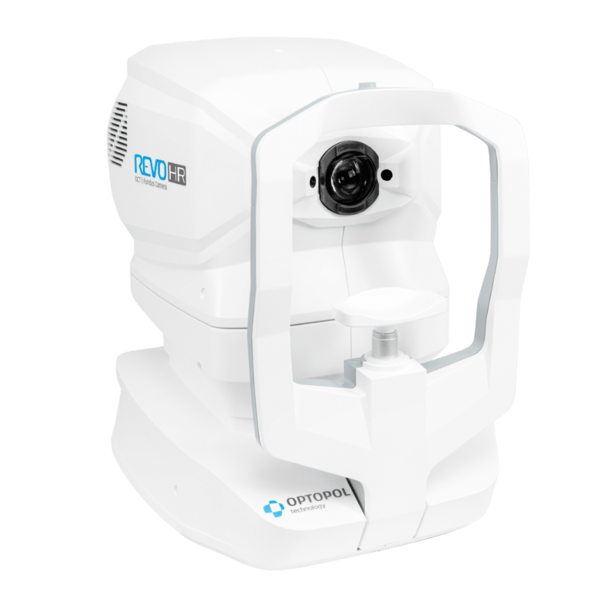
Why Many Clinics in BiH Choose the Optopol REVO Series
The Optopol REVO family (REVO HR, FC130, REVO 80) offers:
-
3 µm tissue resolution
-
130,000 A-scans/s
-
integrated fundus camera (HR, FC130)
-
OCT-A and UWF options
-
full-range scans up to 5.6 mm
-
auto-focus, auto-tracking and auto-capture
-
server and multi-workstation workflow
-
local service and training in BiH
This makes it one of the most advanced and cost-effective solutions in the region.
Conclusion
An OCT eye exam is an essential part of modern ophthalmic diagnostics.
For patients — it is fast, painless and highly informative.
For clinics — choosing the right OCT system significantly improves diagnostic accuracy and service quality.
For OCT demonstrations, device information or quotations, contact us.